Introduction
Infective endocarditis (IE) is a serious infection of the endocardium, the inner lining of the heart chambers and valves. It causes valve damage, formation of vegetations, and systemic embolic events affecting organs such as the brain, lungs, kidneys, and skin. Despite medical advances, IE remains associated with high morbidity and mortality, with a 5-year mortality rate estimated at around 45% [1]. The incidence of IE has increased over recent decades, with rates rising from approximately 10 to nearly 14 cases per 100,000 person-years between 1990 and 2019 [2]. IE most often affects patients with degenerative valve disease, prosthetic valves, or cardiac implantable electronic devices [6,7]. The predominant causative agents are Staphylococcus aureus and viridans group streptococci, but other bacteria, fungi, and rarely viruses can also be responsible [5,8]. Early diagnosis and timely treatment with antibiotics—and surgery when indicated—are crucial to improving patient outcomes.
Clinically, IE can be classified by symptom duration into acute (less than 6 weeks), subacute (6 weeks to 3 months), and chronic (more than 3 months) forms, although this classification is not formally standardized [4].
Methods
This narrative review was conducted through a focused search on PubMed, Scopus, and Web of Science up to 2026. Keywords included “infective endocarditis”, “biomarkers”, “procalcitonin”, “C-reactive protein”, “troponin”, and “natriuretic peptides”. Articles were selected based on their relevance to the diagnostic and prognostic role of biomarkers in IE, including both clinical studies and experimental research on emerging biomarkers. Given the narrative design, this review does not follow a systematic protocol and is thus limited by potential selection bias and heterogeneity in study populations and methods. These factors restrict direct comparison between studies and underline the need for prospective validation in larger cohorts.
Risk factors FOR Infective endocarditis
Risk factors for the development of IE include: degenerative valve diseases, history of invasive procedures in the previous 60 days, congenital valve abnormalities, rheumatic valvular diseases, post-cardiac transplant valvulopathy and the presence of implanted devices [6], [7]. Also, the risk is high in patients with intravenous drug users, immunosuppressed, or with dental procedures and endocarditis in past medical history [7]. The increased use of device placement to improve patient longevity and quality of life has been associated with an enhanced risk of cardiac implantable electronic device (CIED) [9], [10], [11].
The diagnostic of the Infective endocarditis
The diagnosis of IE is established by clinical pictures, blood cultures, echocardiography and other imaging modalities [12].
1. Diagnostic criteria for Infective endocarditis
In accord with the 2023 Duke-International Society for Cardiovascular Infectious Diseases Criteria for Infective Endocarditis (2023 Duke–ISCVID), the clinical diagnosis is determined by combining major and minor criteria (Table no 1):
| Major Clinical Criteria | |
|---|---|
| Microbiological Evidence | Blood cultures positive for typical IE microorganisms (e.g., Staphylococcus aureus, Viridans group streptococci, Enterococcus faecalis, HACEK group, Staphylococcus lugdunensis) isolated from at least 2 separate sets. Note: The 2023 update removed strict time interval constraints between phlebotomies. |
| Microbiological Evidence | Microorganisms typical for intracardiac prostheses only: Microorganisms such as Cutibacterium acnes, Corynebacterium striatum, or Candida spp. represent a major criterion only if the patient has a prosthetic valve or an intracardiac device. |
| Microbiological Evidence | Positive molecular or serological tests for fastidious pathogens (Coxiella burnetii — IgG titer >1:800, Bartonella spp., Tropheryma whipplei) confirmed via PCR, targeted gene sequencing, or metagenomic sequencing. |
| B. Imaging Evidence | Echocardiography (TTE/TEE) or Cardiac CT demonstrating: vegetation, abscess, pseudoaneurysm, intracardiac fistula, valvular perforation, or a new partial dehiscence of a prosthetic valve. |
| B. Imaging Evidence | Abnormal metabolic activity on [18F]-FDG PET/CT within the area of a native valve, prosthetic valve (if implanted > 3 months ago), or along the leads of a cardiac implantable electronic device (CIED). |
| C. Surgical Evidence (New to the 2023 Guidelines) | Direct intraoperative inspection: Direct visualization of a vegetation or abscess by the surgeon during cardiac surgery, even if subsequent laboratory cultures or molecular tests from that tissue return negative. |
| Minor Clinical Criteria | |
| 1. Predisposition | Prosthetic heart valve, prior IE, previous valve repair, congenital heart disease, hypertrophic obstructive cardiomyopathy, or presence of a CIED (pacemaker/defibrillator). Injection drug use (IDU) |
| 2. Fever | Documented body temperature ≥ 38.0°C (100.4°F) |
| 3. Vascular Phenomena | Immune-complex glomerulonephritis, Osler’s nodes, Roth’s spots, or a positive Rheumatoid Factor (RF) |
| 4. Immunological Phenomena | Immune-complex glomerulonephritis, Osler’s nodes, Roth’s spots, or a positive Rheumatoid Factor (RF). |
| 5. Microbiological Evidence | Positive blood cultures that do not meet a major criterion (e.g., a single positive set) or serological/PCR evidence of active infection with an IE-compatible organism from a sterile non-cardiac site. |
| 6. Imaging Evidence | Early abnormal metabolic activity on PET/CT identified within 3 months of a valve or device implantation surgery—does not qualify as a major criterion, but serves as a minor one |
| Source: The 2023 Duke-International Society for Cardiovascular Infectious Diseases Criteria for Infective Endocarditis: Updating the Modified Duke Criteria, Clin Infect Dis. 2023 Aug 22;77(4):518-526. doi: 10.1093/cid/ciad271. | |
Table no 1: Criteria for Infective Endocarditis: Updating the Modified Duke Criteria
2023 Duke–ISCVID diagnosis system [40] use major and minor criteria according to the following scoring system (Figure no 1):
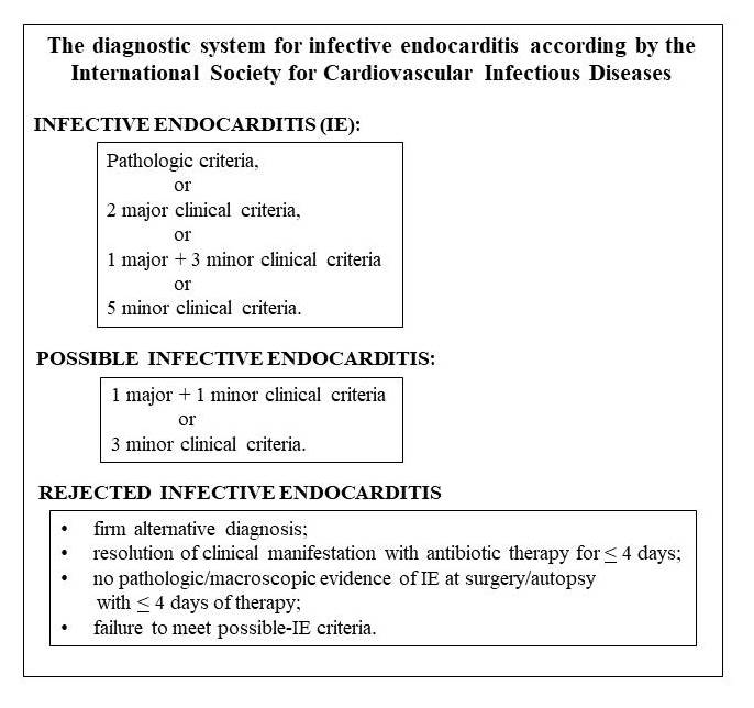
Figure no 1: The diagnostic system for infective endocarditis according by the International Society for Cardiovascular Infectious Diseases
2. Imagistic investigations
2.1. Echocardiography
Echocardiography is a non-invasive diagnostic for IE, which detects the presence of vegetations on heart valves, new valvular lesions and abnormalities in the function and structure of the heart [12], [14].
2.1.1. Transthoracic echocardiography
TTE is a non-invasive ultrasound technique performed by placing a transducer on the chest wall, allowing real-time assessment of cardiac anatomy, valve morphology, chamber size, and function. While it is widely available and often the first imaging modality used, TTE has limited sensitivity in certain patient groups, particularly those with prosthetic heart valves, previous episodes of IE, or poor acoustic windows resulting from obesity, chest wall deformities, or lung disease. In these patients, negative or inconclusive TTE results necessitate further imaging by transesophageal echocardiography [13,14].
2.1.2. TEE (Transesophageal echocardiography)
TEE involves inserting a specialized ultrasound probe into the esophagus, placing it in close proximity to the heart and providing superior image quality. This technique offers higher resolution and approximately 95% sensitivity for detecting intracardiac vegetations and complications, making it the gold standard when TTE is negative or inconclusive, or when prosthetic valve endocarditis and device-related infections are suspected [18]. TEE is particularly useful for detecting valvular and perivalvular complications such as abscesses, pseudoaneurysms, and fistulas [15].
2.2. Additional Imaging Modalities
2.2.1. Cardiac Computed Tomography (CT)
Cardiac CT has proven to be a good exploration technique when a better depiction of valvular complications is needed or when echocardiography proves to be insufficient in both native and prosthetic valve endocarditis [16].
2.2.2. Positron Emission Tomography/Computed Tomography (PET/CT)
PET/CT is increasingly utilized in IE diagnosis, especially for prosthetic valve endocarditis and infections of cardiac implantable devices. The 2023 European Society of Cardiology (ESC) Guidelines recommend PET/CT for patients with suspected prosthetic valve or device-related IE where echocardiography and blood cultures are inconclusive, particularly when the prosthesis was implanted over three months earlier. Abnormal fluorodeoxyglucose uptake on PET/CT is now included as a major criterion in the diagnosis of prosthetic valve endocarditis [17,18]. Rouzet et al. demonstrated the added value of PET/CT in improving diagnostic accuracy in these cases [19]. The particularities and efficacy of the mains imaging techniques used in the diagnosis of infective endocarditis and of their complications, are presented in the Table no 2.
Table no 2: Imaging techniques used in the diagnosis of infective endocarditis (adapted from Guillermo Cuervo, et al: Current Challenges in the Management of Infective Endocarditis)
| Imaging techniques used in the diagnosis of infective endocarditis and of their complications | |
|---|---|
| Transthoracic echocardiography (TTE) | Mainstay of diagnosis. Limited sensitivity in prosthetic valve endocarditis and paravalvular complications. |
| Transesophageal echocardiography (TEE) | Mainstay of diagnosis. Indicated in prosthetic valve endocarditis and for the detection and characterization of valvular and paravalvular complications. |
| Three-dimensional transesophageal echocardiography (3D-TEE) | Complement to TEE when characterizing valvular and paravalvular complications. Surgical planning. |
| Intraoperative transesophageal echocardiography | Routine exploration when surgery is performed. Assessment of post-operative anatomy and ventricular function. |
| Cardiac computed tomography (CT) | Better characterization of complications involving an aortomitral fibrous body and the aortic root. Alternative to coronary angiography for exploration in patients with aortic endocarditis. |
| Positron emission tomography /computed tomography (PET/CT) | Major Duke criteria for diagnosis of prosthetic valve endocarditis and device-associated endocarditis. Increasing use in infective endocarditis associated with TAVI and LVAD. Detection of extracardiac complications of endocarditis or alternative sources of infection. |
| Radiolabeled white blood cell single-photon emission computed tomography/computed tomography (WBC-SPECT/CT) | Complement to PET/CT when results are non-conclusive. Similar performance to that of PET/CT. |
| Brain magnetic resonance imaging (MRI) | Detection and characterization of silent or clinically evident intracranial complications. Evaluation of evolving changes in intracranial complications that may influence the timing for surgery. |
| Thoracoabdominopelvic computed tomography | Not recommended as a routine exploration in asymptomatic patients. |
| Contrast-enhanced ultrasound | Promising results on detecting intraabdominal emboli as an alternative to CT. |
| Colonoscopy | Detection of colorectal disease or neoplastic processes in Streptococcus gallolyticus or Enterococcus faecalis endocarditis. |
Biomarkers for the Diagnosis of Infective Endocarditis
Biomarkers are measurable biological indicators that reflect normal physiological processes, pathological changes, or responses to therapeutic interventions. They play a central role in disease detection, in prognosis and monitoring of treatment effectiveness.
1. C-reactive protein (CRP)
C-reactive protein (CRP) is an acute-phase reactant synthesized predominantly by hepatocytes [19] in response to pro-inflammatory cytokines, particularly interleukin-6 (IL-6). The protein plays a role in the innate immune response by binding to phosphocholine on microbial surfaces and damaged host cells, activating the complement system and facilitating phagocytosis. CRP is a key inflammatory marker in infective endocarditis and is used for diagnosis support, monitoring disease activity, and prognosis.
CRP is a non-specific marker that can increase due to many infectious or non-infectious diseases, and which has not been accepted as a diagnostic criterion [21]. Increases in CRP values were noted at diagnosis of the disease and were correlated with disease activity and severity. Also, persistence of elevated or rising levels suggests an inadequate response to antimicrobial therapy or the development of complications (heart failure, embolic events, perivalvular abscesses) and an increased risk of mortality [22]. Serial measurement of CRP during antimicrobial therapy provides valuable information regarding treatment efficacy. Higher CRP values are frequently observed in acute IE, particularly in infections caused by Staphylococcus aureus, compared with subacute forms associated with less virulent organisms [23]. CRP should be measured at least weekly during the treatment of patients with IE [22].
2. D-dimer
D-dimer is a protein fragment that is released into the bloodstream as a result of the dissolution of blood clots [24], [8]. Several studies (Lin et al., 2021; Mitrosinka et al., 2021; Turak et al., 2017) have demonstrated that D-dimer levels are significantly elevated in patients with infective endocarditis (IE), reflecting activation of coagulation and fibrinolytic pathways secondary to systemic inflammation and valvular vegetation formation [2]. In infective endocarditis, D-dimer levels are often elevated, but they are not specific and not diagnostic. They can also be detected in other conditions such as deep vein thrombosis or disseminated intravascular coagulation [25]. Elevated D-dimer values are associated with an increased risk of thromboembolic complications and a poor prognostic. Outcome of recent clinical studies and trials have shown a correlation between high values of D-dimer levels at-admission and risk of in-hospital complications and death in patients with endocarditis. [26].
3. NT-proBNP
NT-proBNP (N-terminal pro-B-type natriuretic peptide) is an important marker of cardiac stress and prognostic in infective endocarditis, that is not included in the modified Duke diagnostic criteria. The elevated levels reflect hemodynamic stress caused by infection, myocardial dysfunction and disease severity. NT-proBNP is often used in the diagnosis and monitoring of heart failure and treatment response [27, 28]. Natriuretic peptides (NPs), including B-type NP (BNP) and the N-terminal fragment of its prohormone (NT-proBNP), are endogenous cardiac hormones released primarily from ventricular myocytes in response to myocyte stretch. Also, increased levels of NT-proBNP can be found in pulmonary embolism and acute respiratory distress syndrome [8], [29]. Determination of elevated NT-proBNP levels at admission or during hospitalization have been associated with complicated disease courses and poorer prognosis. Studies show that there are no significant differences between NT-proBNP-levels in patients with native valve and those with prosthetic valve in endocarditis. [27], [28].
4. Procalcitonin (PCT)
Procalcitonin (PCT) is a precursor to the hormone calcitonin, which is released in response to endotoxins and systemic mediators released during bacterial infections. Procalcitonin (PCT) is the protein given most attention in the last decade as a putative biomarker for IE [5]. The values of the procalcitonin were an important diagnostic marker in EI, which increases during several bacterial infection and decreases if the infection improves under treatment. Plasma level of PCT rises within 3–6 h from initial clinical manifestation of sepsis and decreases when severe infection is controlled under therapy [41]. Monitoring PCT levels is an assessment tool for guiding therapeutic decisions regarding the duration of antimicrobial treatment. High PCT concentrations are typically found in bacterial infections, whereas lower levels predominate in viral infections [30]. Jereb et al. found highest PCT levels in patients with Staphylococcus aureus endocarditis [31]. Procalcitonin levels were initially indicated to assess the risk of critically ill patients for progression to severe sepsis and shock and for the cumulative risk of all cause mortality at 28 days after admission. Elevated PCT concentrations can also be found in the absence of infection after major surgery, trauma, severe burns, prolonged cardiogenic shock, chronic kidney diseases, autoimmune or malignant diseases [32].
5. Troponins
Troponins are a group of biomarkers widely utilized in diagnosing and managing acute coronary syndromes, particularly myocardial infarction. Cardiac troponin is frequently elevated in patients with infective endocarditis (IE). Troponin I and troponin T are highly sensitive and specific biomarkers of myocardial injury. High levels of troponin in infective endocarditis reflect myocardial injury from embolic, inflammatory, or hemodynamic mechanisms. Elevated troponin (Tn) levels are very common in infective endocarditis (IE), occurring in up to 84% of cases, and serve as a critical, independent marker for poor prognosis [33]. Multiple observational studies demonstrate that elevated troponin levels in IE are associated with higher mortality rates, long-term hospitalization, risk of intracardiac complications (peri-annular abscess) and increased need for surgical intervention [33], [34]. Elevated troponin levels are found in other conditions such as pulmonary embolism, sepsis and renal failure.
6. Cystatin C
Cystatin C (Cys C) is a biomarker of kidney function. Cystatin C is a 13-kDa cysteine protease inhibitor, freely filtered at the glomerulus, minimally affected by age, sex, and muscle mass. Studies indicate that CysC is a strong predictor of 90-day and 5-year mortality in IE patients [42]. CysC also offers superior ability to diagnose acute kidney injury and declining GFR compared with creatinine [8]. Renal dysfunction in infective endocarditis can result from several mechanisms, including:
1. immune-complex glomerulonephritis;
2. renal embolization;
3. sepsis and renal hypoperfusion;
4. heart failure and cardiorenal syndrome;
5. therapy with nephrotoxic antimicrobial drugs.
7. Interleukin-6 (IL-6)
IL-6 is a pro-inflammatory cytokine produced by various cells, (macrophages and endothelial cells), being one central mediator of the systemic inflammatory response in infective endocarditis. IL-6 reflects the intensity of the host immune response to valvular infection and represents a marker of disease activity [3]. Serum interleukin-6 (IL-6) level can be used to aid in diagnosis of infective endocarditis (IE), especially when blood cultures are negative [3].
8. Serum calcium level
Serum calcium level represents another biomarker used in detection of IE. Clinical studies suggest that low serum calcium levels may be associated with severe infection (indicated by elevated inflammatory parameters), organ dysfunction, and increased in-hospital mortality [8]. The calcium deposition on the valve surface can be detected by echocardiography and is associated with an increased risk of embolic complications [36].
9. S100A11 (Calcium-Binding Protein S100A11)
S100A11 (Calcium-Binding Protein S100A11) is a calcium-binding protein from the S100 family. In patients with infective endocarditis (IE) represents a biomarker of inflammation and endothelial injury [3]. The increased expression of the S100A11 gene was essentially related to Staphylococcus infection.
10. Aquaporins (AQP9)
Aquaporins (AQP9) is expressed in multiple tissues, including the liver, spleen, brain and plays a role in transplant of glycerol and other small solutes [37]. In IE patients, the upregulation of the aquaporin-9 gene (AQP9) is associated with the occurrence of acute heart failure. The detection of predictors of acute heart failure is of crucial importance because it can indicate the need for valvular surgery at an early stage of the disease [38].
Future biomarkers for infective endocarditis (IE)
Future biomarkers for infective endocarditis (IE) focus on improving early diagnosis, risk stratification, and predicting complications (heart failure, embolic event). Table no 3 shows the main biomarkers used and studied in several recent clinical trials, who assured transition from generic systemic inflammatory markers to integrated proteomic signatures.
Table no 3: The main investigational biomarkers of infective endocarditis
| Characteristics | Mechanism | Indications for Use as potential markers | Relevant experimental studies | |
|---|---|---|---|---|
| LRG1 (Leucine-Rich Alpha-2-Glycoprotein 1) | A novel serum protein linked to inflammation-driven angiogenesis. | It modulates TGF-beta signaling pathways, accelerating abnormal neovascularization inside growing bacterial vegetations, making them structurally unstable. | Recomended to distinguish true IE from non-endovascular sepsis or sterile systemic inflammation. | Zhang et al., Nature Communications (2025): Proteomic profiling of plasma and vegetation samples from 238 IE patients and 100 controls (validated across external plasma cohorts) identified LRG1 as a potential biomarker significantly associated with infection severity and neutrophil activity |
| NDUFB4 (NADH Dehydrogenase Subunit) | A metabolic tissue-and-plasma marker discovered through spatial proteomic profiling of surgically excised valvular vegetations. | Reflects severe mitochondrial dysfunction and oxidative exhaustion of local macrophages trying to breach the protective bacterial biofilm of the vegetation. | Assessing the deep structural destruction of perivalvular tissue and stratifying patients at high risk for myocardial fistulization or sudden conduction blocks | Integrated plasma and vegetation proteomics, Nature Communications (2025): Advanced mass spectrometry network analysis identified NDUFB4 as a metabolic clearance and inflammation biomarker critically tied to adverse structural events and glycometabolism dysfunction in severe IE. |
| Host Immune Hub Genes (ITGAM, FCGR3B, FCGR3A) | based on measuring messenger RNA (mRNA) expression levels | These hub genes code for essential leukocyte integrins and Fc-gamma receptors necessary for white blood cell adhesion and the clearance of circulating immune complexes trapped in cardiac lesions. | Highly specific differential diagnosis in complex subacute cases mimicking autoimmune conditions, particularly when secondary to chronic periodontal infections. | Bioinformatics and Host Transcriptomics in Sepsis & Carditis (2022–2024): RNA-seq data networks confirm these core "hub genes" orchestrate neutrophil activation, tissue adhesion, and macrophage dynamics during heavy endovascular biofilm infection and early sepsis. |
| MR-proADM (Mid-Regional Pro-Adrenomedullin) | A stable, reliable plasma peptide that acts as an direct indicator of systemic endothelial barrier failure. | Released by endothelial cells in response to mechanical shear stress and bacterial invasion to protect vascular integrity and modulate local vascular tone [38]. | Monitoring patients during the early post-operative window following valve replacement surgery to differentiate sterile systemic inflammatory response syndrome (SIRS) from early prosthetic valve endocarditis (PVE) or dehiscence. | Montrucchio et al., International Journal of Molecular Sciences (2025): Clinical case series of post-cardiopulmonary bypass patients demonstrating that MR-proADM significantly increases in active post-surgical infective endocarditis (median 5.9 ng/mL) compared to standard elective cardiac surgery (median 1.1 ng/mL). |
| SeptiCyte RAPID | PCR based test for sepsis who generate numeric likelihood score (SeptiScore) within an hour. | Quantifies the expression ratios of 4 distinct host genes directly from a peripheral blood sample, distinguishing infectious immune activation from sterile post-surgical inflammation | Emergency differentiation of active IE from sterile cardiotomy shock or post-bypass inflammation in patients presenting with acute post-operative fevers. | University of Turin Hospital Case Series / MDPI (2025): Evaluated in cardiosurgical intensive care units, demonstrating that this FDA-cleared host-response assay successfully differentiates acute bacterial activation from sterile post-bypass systemic inflammation. |
CONCLUSIONS
The determination of circulating biomarkers may represent a valuable adjunctive tool in the diagnostic workup and clinical management of infective endocarditis. Imagistic investigation (echocardiography transthoracic or transesophageal) and blood cultures remain the gold standard for diagnosis. The use of biological markers (such as CRP, procalcitonin, or troponins) provides critical tools for early risk stratification, monitoring therapeutic response, and detecting major complications. These biological indicators redefine the prognosis of critically ill patients. Elevated on-admission D-dimer levels have been observed to correlate with a higher risk of in-hospital adverse events and mortality, suggesting a potential role in prognosis. Furthermore, cardiac-specific biomarkers, including high-sensitivity troponins and natriuretic peptides (BNP/NT-proBNP), may provide useful early indicators of subclinical myocardial injury, structural valve deterioration, or impending hemodynamic failure.